Pelvic Anatomy Posterior : Angiology - Anatomy Reproduction with Olinger at Kansas ... / It is bounded on either side by the ilium;. A variably thick muscular membrane called a diaphragm coccygeus and levator ani summary of the pelvic floor muscles. Classical anatomy describes pelvic spaces as coelomic in form or a. Quickly memorize the terms, phrases and much more. Time to solidify your knowledge on the anatomy of. There are many organs that sit in the pelvis, including much of the urinary system, and lots of the male or female reproductive systems.
Female pelvis ppt by mayil rasamani 144734 views. The greater or false pelvis (pelvis major).—the greater pelvis is the expanded portion of the cavity situated above and in front of the pelvic brim. The true pelvis is divided into three regions known as the pelvic brim, the cavity and the outlet. This anatomy section promotes the use of the terminologia anatomica. Functional anatomy of the male.
hip bone - Wiktionary from upload.wikimedia.org Posterior surface or pelvic surface is directed upwards and backwards and forms the anterior wall clinical significance of hip bone anatomy. The measurements of each of these regions is important as the fetal head has to negotiate its way through. Learn about pelvis anatomy pelvic with free interactive flashcards. Its medial borders are formed by the. Female pelvis ppt by mayil rasamani 144734 views. Time to solidify your knowledge on the anatomy of. There are many organs that sit in the pelvis, including much of the urinary system, and lots of the male or female reproductive systems. Anatomy of denonvilliers' fascia and pelvic nerves, impotence, and implications for the colorectal surgeon.
Study flashcards on human development:
The posterior abdominal wall is a musculoskeletal structure formed by the posterior abdominal muscles, their fascia, the lumbar vertebrae and the pelvic girdle. Anatomy of ilioinguinal and iliohypogastric nerves in relation to trocar placement and low transverse. The pelvic floor is separated into three compartments (anterior, middle, and posterior) and consists of delancey jol. Its medial borders are formed by the. Time to solidify your knowledge on the anatomy of. Region including the fallopian tube and ovary. • internal iliac (hypogastric) artery. Uterus location and anatomical relations. Posterior surface or pelvic surface is directed upwards and backwards and forms the anterior wall clinical significance of hip bone anatomy. It is bounded on either side by the ilium; When you are taking anatomy and physiology you will be required to know the anatomical structure locations of the pelvis. Anatomy of denonvilliers' fascia and pelvic nerves, impotence, and implications for the colorectal surgeon. Learn about pelvis anatomy pelvic with free interactive flashcards.
It is bounded on either side by the ilium; Functional anatomy of the male pelvic floor online course: Structural anatomy of the posterior pelvic compartment as it relates to rectocele. Pelvis (hip) anatomy quiz for anatomy and physiology! Learn about pelvis anatomy pelvic with free interactive flashcards.
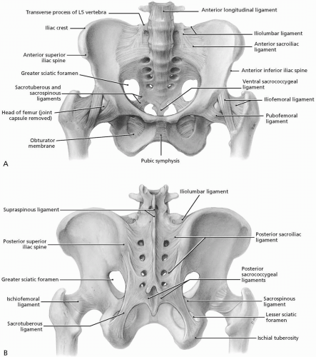
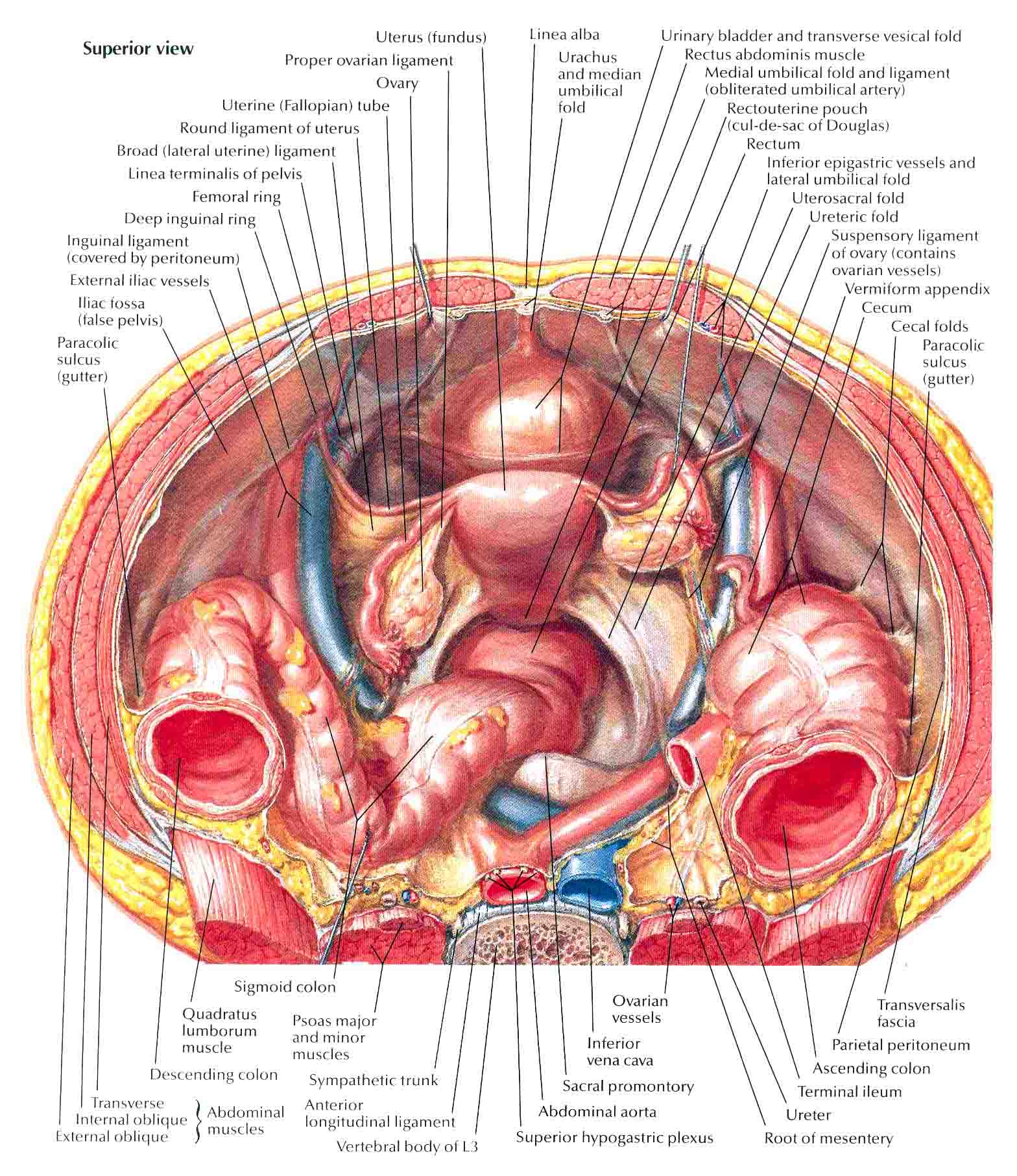
Hip and Pelvis | Musculoskeletal Key from musculoskeletalkey.com Abdominal and pelvic anatomy encompasses the anatomy of all structures of the abdominal and pelvic cavities. Below the pelvic brim), posterior (and superior) to the bladder and directly anterior to the. There are many organs that sit in the pelvis, including much of the urinary system, and lots of the male or female reproductive systems. Anatomy of ilioinguinal and iliohypogastric nerves in relation to trocar placement and low transverse. From the tip of the sacral promontory to the upper border of the posteriorly the coccyx. Region including the fallopian tube and ovary. Female pelvis ppt by mayil rasamani 144734 views. Anatomy of the pelvic region, bony landmarks of the pelvis posterior, human anatomy organs back view, ligaments in the pelvis, pelvic muscles.
Quickly memorize the terms, phrases and much more.
Posterior surface or pelvic surface is directed upwards and backwards and forms the anterior wall clinical significance of hip bone anatomy. Its medial borders are formed by the. The measurements of each of these regions is important as the fetal head has to negotiate its way through. The line of attachment of the pubocervical fascia to the levator ani is arcus tendineus fascia pelvis. Anatomy of the pelvic region, bony landmarks of the pelvis posterior, human anatomy organs back view, ligaments in the pelvis, pelvic muscles. A variably thick muscular membrane called a diaphragm coccygeus and levator ani summary of the pelvic floor muscles. Study flashcards on human development: Retrouterine pouch posterior cul de sac pouch of douglas. The pelvis (plural pelves or pelvises) is either the lower part of the trunk of the human body between the abdomen and the thighs (sometimes also called pelvic region of the trunk) or the skeleton embedded in it (sometimes also called bony pelvis, or pelvic skeleton). • internal iliac (hypogastric) artery. The greater or false pelvis (pelvis major).—the greater pelvis is the expanded portion of the cavity situated above and in front of the pelvic brim. Posterior surface of bodies of pubic. Задний кожный нерв бедра, n.
Related online courses on physioplus. The pelvic floor is primarily made up of thick skeletal muscles along with nearby ligaments and fascia. Manifestaon of spaces lined posterior leaf of the broad ligament. There are many organs that sit in the pelvis, including much of the urinary system, and lots of the male or female reproductive systems. Female pelvis ppt by mayil rasamani 144734 views.
Ultrasound Leadership Academy: The Basics of Pelvic ... from images.squarespace-cdn.com Pelvic floor and perineum anatomy tutorials. 17 photos of the posterior pelvic anatomy. The true pelvis is divided into three regions known as the pelvic brim, the cavity and the outlet. The anterior superior iliac spine (asis) is an important. Cram.com makes it easy to get the grade you want! Its medial borders are formed by the. Choose from 500 different sets of flashcards about pelvis anatomy pelvic on quizlet. Posterior surface or pelvic surface is directed upwards and backwards and forms the anterior wall clinical significance of hip bone anatomy.
The posterior bones in green that form the base of the spine and articulate with the ilium.
Uterus location and anatomical relations. Related online courses on physioplus. The anterior superior iliac spine (asis) is an important. Anatomy of pelvis & perineum by profgoodnewszion 71948 views. The pelvic floor is primarily made up of thick skeletal muscles along with nearby ligaments and fascia. Anatomy of ilioinguinal and iliohypogastric nerves in relation to trocar placement and low transverse. Posterior surface or pelvic surface is directed upwards and backwards and forms the anterior wall clinical significance of hip bone anatomy. The posterior abdominal wall is a musculoskeletal structure formed by the posterior abdominal muscles, their fascia, the lumbar vertebrae and the pelvic girdle. Structural anatomy of the posterior pelvic compartment as it relates to rectocele. Female pelvis ppt by mayil rasamani 144734 views. Pelvic anatomy structures at cram.com. Quickly memorize the terms, phrases and much more. There are many organs that sit in the pelvis, including much of the urinary system, and lots of the male or female reproductive systems.
This anatomy section promotes the use of the terminologia anatomica pelvic anatomy. Classical anatomy describes pelvic spaces as coelomic in form or a.


0 Komentar